About me
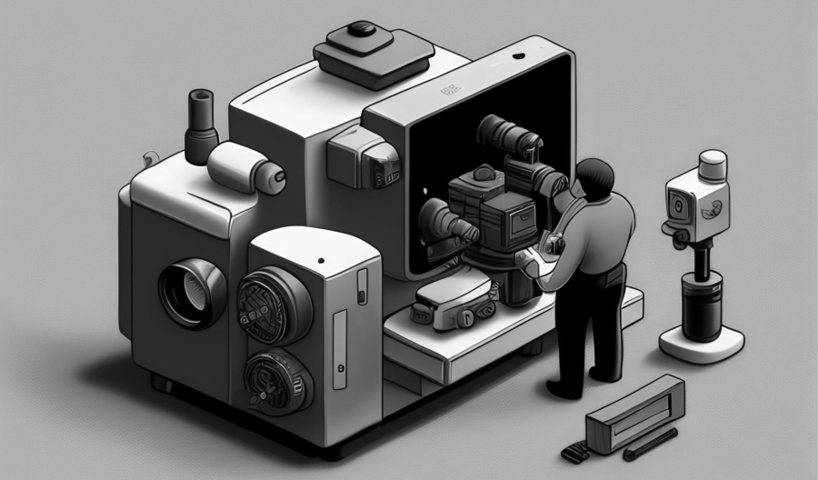
Hi there! I’m an Imaging and Bioimage Informatics Specialist at the Imaging Core of the Lerner Research Institute (LRI), Cleveland Clinic, where I’ve been working since 2018. In this role, I have extensively utilized light microscopy techniques such as confocal microscopy, total internal reflection fluorescence (TIRF) microscopy, multi-photon microscopy, and multi-spectral imaging to support a variety of biomedical research projects. Additionally, I have leveraged my experience with electron microscopy, including both Scanning Electron Microscopy (SEM) and Transmission Electron Microscopy (TEM), to further support biomedical research at LRI.
Collaborating with researchers and enhancing our image processing capabilities has been incredibly rewarding. I started with commercial tools like Image Pro and HALO, and expanded to open-source tools like ImageJ, QuPath, CellProfiler, and Python. My projects include analyzing whole slide images and developing pipelines for immuno-oncology research, where these imaging techniques have been crucial in studying cellular interactions and phenotyping cells.
I am leading the efforts to establish the Bioimage Analysis Core at LRI, providing cutting-edge tools and expertise. Leveraging deep learning and AI, I’ve enhanced the accuracy of our image analysis, overcoming the limitations of traditional methods. My technical acumen and passion for imaging drive me to push the boundaries of what’s possible and support groundbreaking biomedical research.
As part of my responsibility to implemente iLab for the Imaging Core, I developed software that verifies a user’s identity and accounting information, tracks equipment usage, and facilitates Imaging Core billing in iLab. I have also developed a plug-in for the software package Digital Micrograph, which allows our electron microscope operators to verify and correct patient information for the clinical diagnostic images.
Before joining the Imaging Core of Lerner Research Institute at Cleveland Clinic, I performed my dissertation research at the Advanced Chemical Imaging Facility in the laboratory of Dr. John F. Turner II within the Department of Chemistryat Cleveland State. As part of this work, I have developed a novel broadly tunable surface plasmon-coupled wavelength filter for visible and near infrared hyperspectral imaging. The novel filter technology is designed to minimize the short coming of the current wavelength filter technologies for wide-field hyperspectral imaging like the liquid crystal tunable filter (LCTF) and acousto-optic tunable filter (AOTF). In addition, the surface plasmon filter that I developed is rugged, compact, and able to be integrated into existing optical microscopes and handheld imagers. Also as part of my dissertation work, I have constructed a wide-field AOTF Raman imaging system to investigate the distribution of crystalline and amorphous domains in an important biodegradable implant material, poly-L-lactide. In its crystalline state, poly-L-lactide exhibits a greater elastic modulus, similar to bone, and is piezoelectric. The Raman imaging modality can discern subtle variations in the crystalline content of poly-L-lactide nondestructively and is useful for characterizing biomaterials in general. Both research projects required considerable effort in optical design, photonic device construction, optical testing, computer interfacing to control acquisition, computer programming (Visual Basic, Visual C++, MatLab, etc.), and novel multivariate methods for data analysis.
In addition to my dissertation research, I also have considerable experience in scanning probe methods like atomic force microscopy (AFM), scanning electron microscopy (SEM) and energy dispersive x-ray spectroscopy (EDS). In fact, as part of my teaching assistantship, I was assigned to oversee the SEM lab and to provide SEM analysis and training on the SEM/EDS systems. Before initiating my Ph.D. studies at Cleveland State University, I conducted research at Governors State University where I gained significant experience using chromatography for extraction, identification and quantification of trace amounts of contaminants in foods. My work there was focused on the detection of melamine and its derivatives in milk and other food products employing LC/MS and GC/MS.